Zangwill LM, Bowd C, Berry CC, Williams J, Blumenthal EZ, Sanchez-Galeana CA, Vasile C, Weinreb RN.
Archives of Ophthalmology 2001;119(7):985-993
Glaucoma Center, Department of Ophthalmology, University of California-San Diego, La Jolla, CA 92093-0946, USA.
ABSTRACT:
OBJECTIVE: To compare the ability of 3 instruments, the Heidelberg Retina Tomograph (HRT), the GDx Nerve Fiber Analyzer (GDx), and the Optical Coherence Tomograph (OCT), to discriminate between healthy eyes and eyes with early to moderate glaucomatous visual field loss.
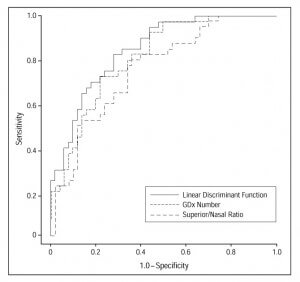
SUBJECTS AND METHODS: Forty-one patients with early to moderate glaucomatous visual field loss and 50 healthy subjects were included in the study. The HRT, GDx, and OCT imaging and visual field testing were completed on 1 eye from each subject within a 6-month interval. Statistical differences in sensitivity at fixed specificities of 85%, 90%, and 95% were evaluated. In addition, areas under the receiver operating characteristic (ROC) curve were compared.
RESULTS: No significant differences were found between the area under the ROC curve and the best parameter from each instrument: OCT thickness at the 5-o’clock inferior temporal position (mean +/- SE, 0.87 +/- 0.04), HRT mean height contour in the nasal inferior region (mean +/- SE, 0.86 +/- 0.04), and GDx linear discriminant function (mean +/- SE, 0.84 +/- 0.04). Twelve HRT, 2 GDx, and 9 OCT parameters had an area under the ROC curve of at least 0.81. At a fixed specificity of 90%, significant differences were found between the sensitivity of OCT thickness at the 5-o’clock inferior temporal position (71%) and parameters with sensitivities less than 52%. Qualitative assessment of stereophotographs resulted in a sensitivity of 80%.
CONCLUSION: Although the area under the ROC curves was similar among the best parameters from each instrument, qualitative assessment of stereophotographs and measurements from the OCT and HRT generally had higher sensitivities than measurements from the GDx.
Figures from this article:


